
3D Roughness Analysis Scanning Electron Microscope ERA-600 Series
【To customers considering the ERA series】
Notice of Order Suspension
3D Roughness Analysis Scanning Electron Microscope
ERA-600 Series
- Measurement and Analysis Equipment
topography measurement, and roughness analysis
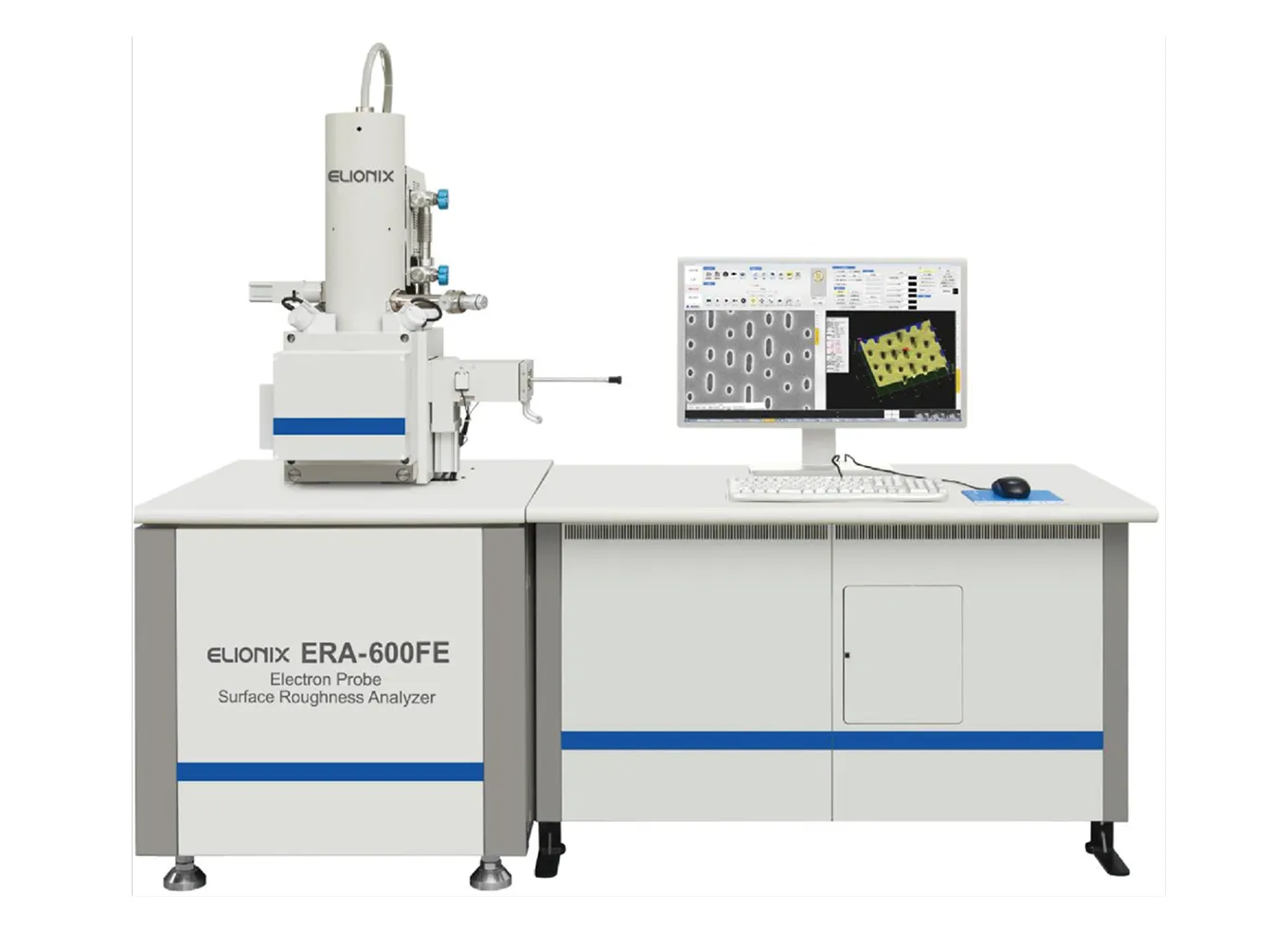
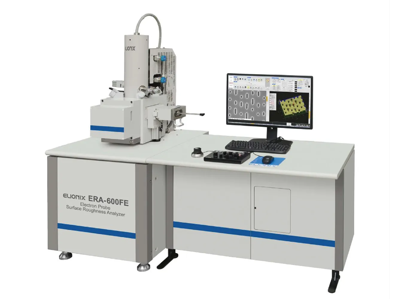
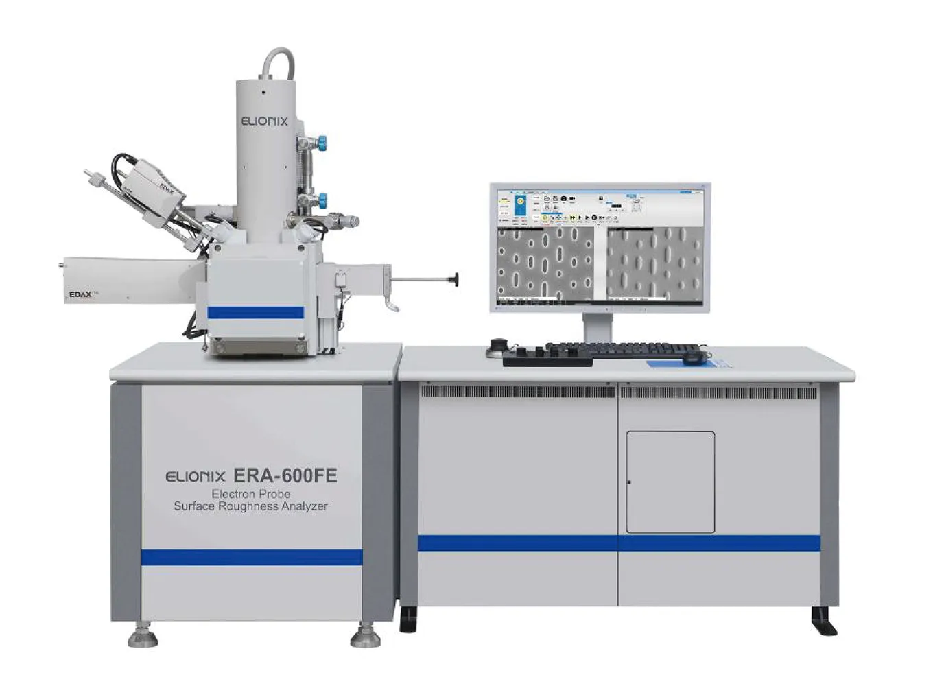
Key Features
Field-Emission Scanning Electron Microscope for Three-Dimensional Roughness Analysis
The definitive choice for high-precision, high-resolution surface profilometers.
This single instrument enables high-resolution SEM observation, surface profilometry, and the calculation of surface property parameters!
Recipient of the Japan Society for Precision Engineering Technical Award.
What can you do with the ERA series? What can you learn from it?
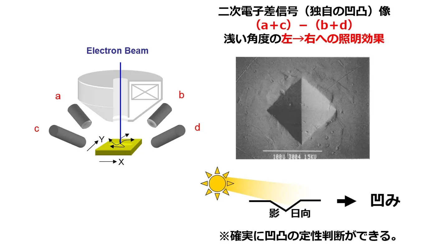
By removing compositional information, the image is reduced to a representation of topographical contrast alone.
Unlike standard SEM images, where topographical details are often ambiguous, this image allows for reliable determination of topographical features and enables precise shape measurement.
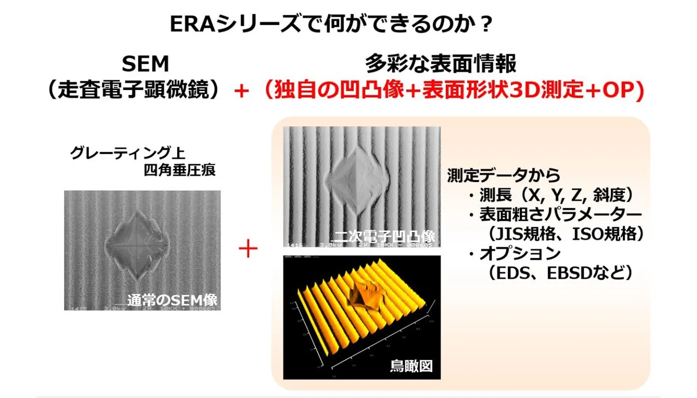
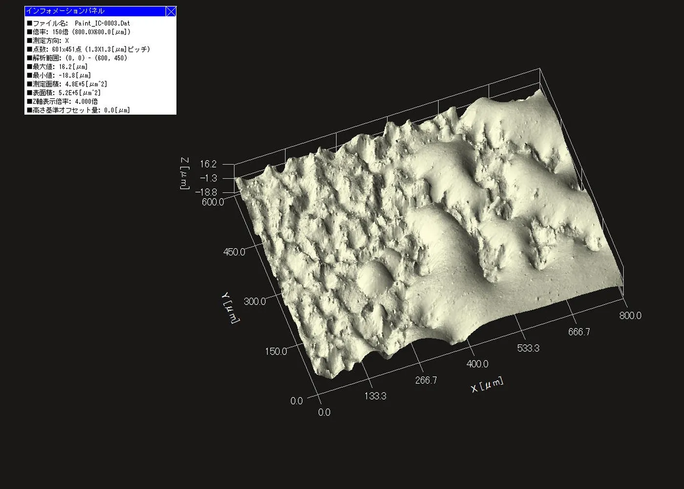
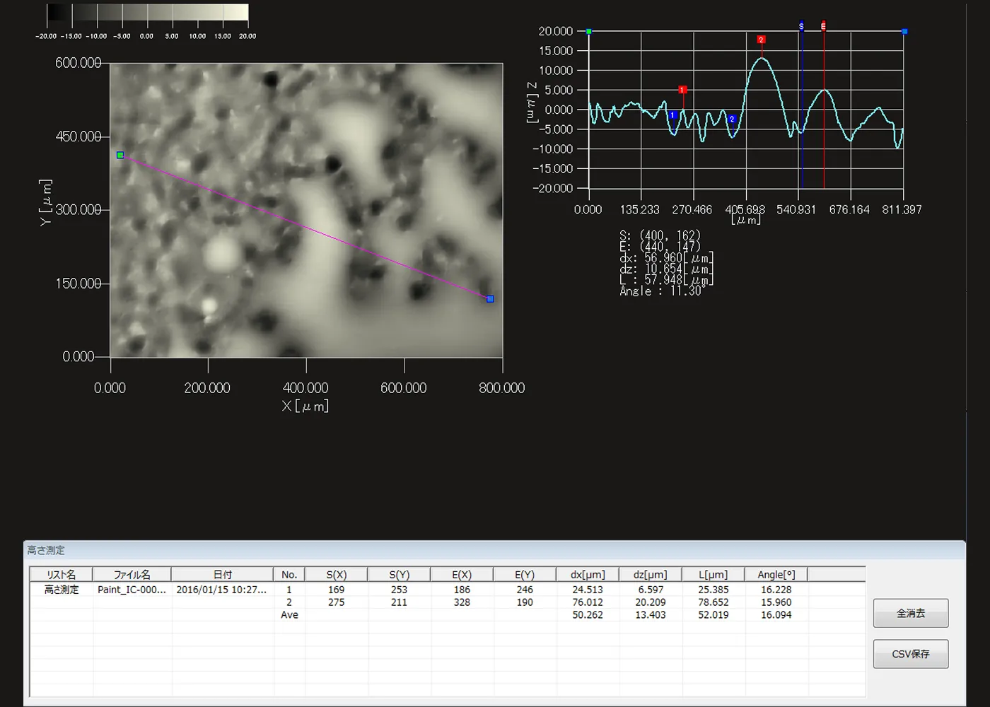
Various surface property parameters can be obtained
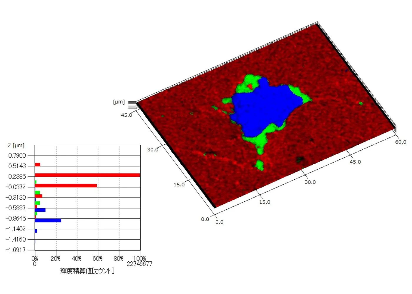
- You can obtain 3D bird's-eye-view images, contour maps, and surface texture parameters compliant with JIS and ISO standards.
- By linking these SEM images and 3D measurement data with Windows-compatible file server software, you can manage your data efficiently. This also significantly improves the efficiency of creating papers and reports.
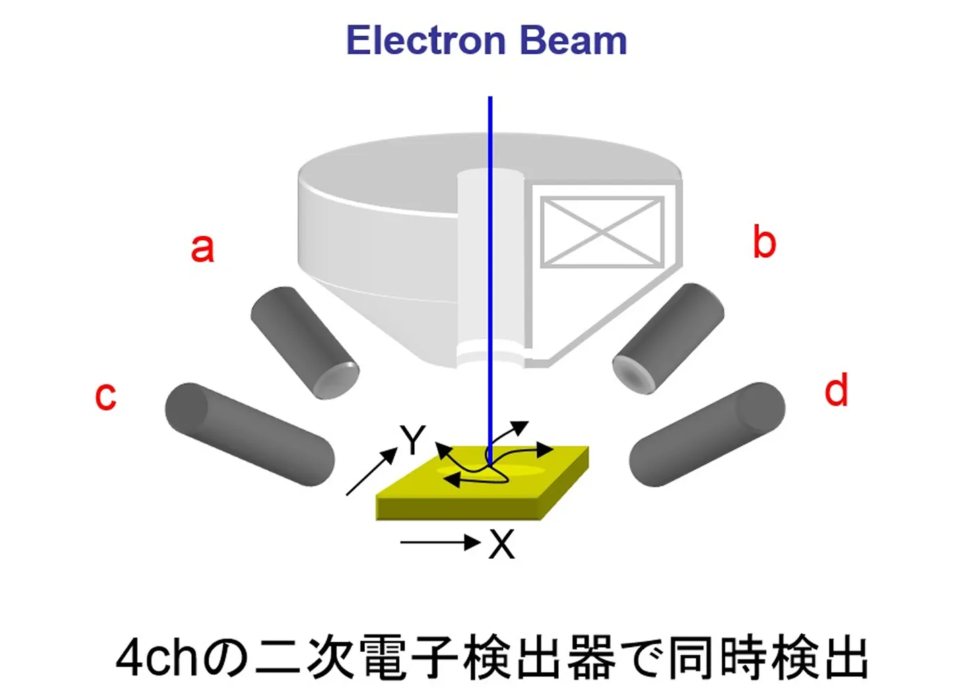
The world's only 3D imaging system equipped with four secondary electron detectors
Additionally, options such as EDS, EBSD, and CL can be installed.
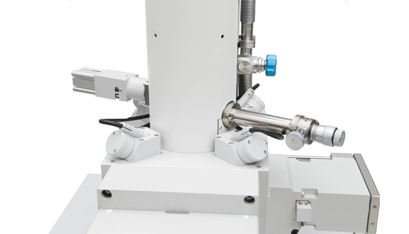
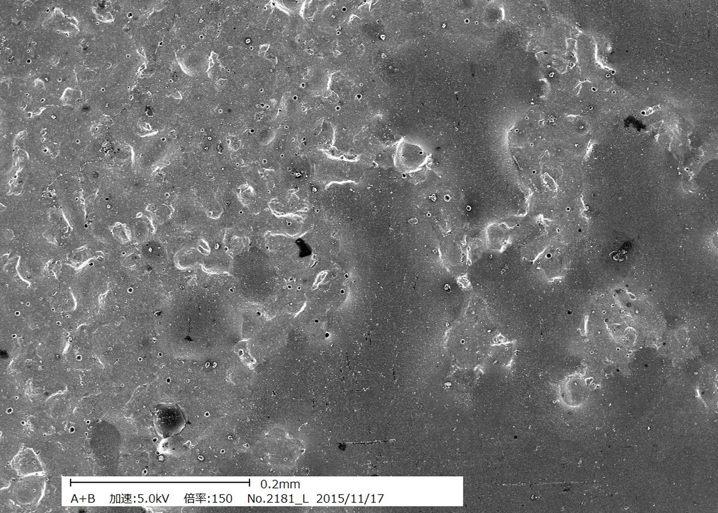
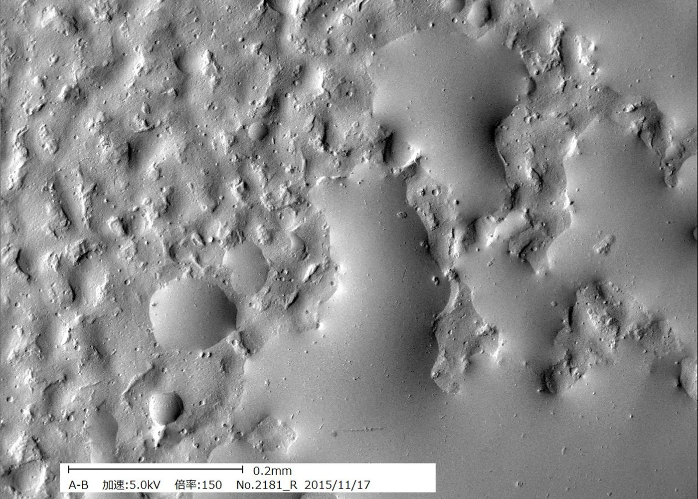
High resolution in both the vertical and horizontal directions
High usability
- User-friendly Japanese menus and mouse-based operation.
- Equipped with an auto function for easy operation. In addition, the use of an integration unit provides clear, real-time still images, enabling observation and measurement even in brightly lit rooms.
Minimal damage to the sample
- Because the electronic probe scans the sample surface at high speed to take measurements, it does not damage the sample surface.
- The irradiation current is also extremely low, at 10 pA or less, allowing us to measure even samples that are susceptible to beam damage.
Easy to maintain
Because it is equipped with a proprietary long-life electron source (Schottky type), it requires virtually no routine maintenance.
3D Source Mapping with EDS Option Installed
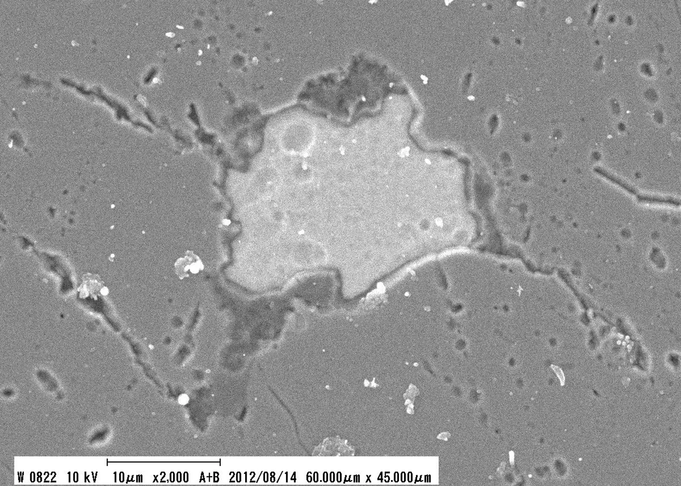
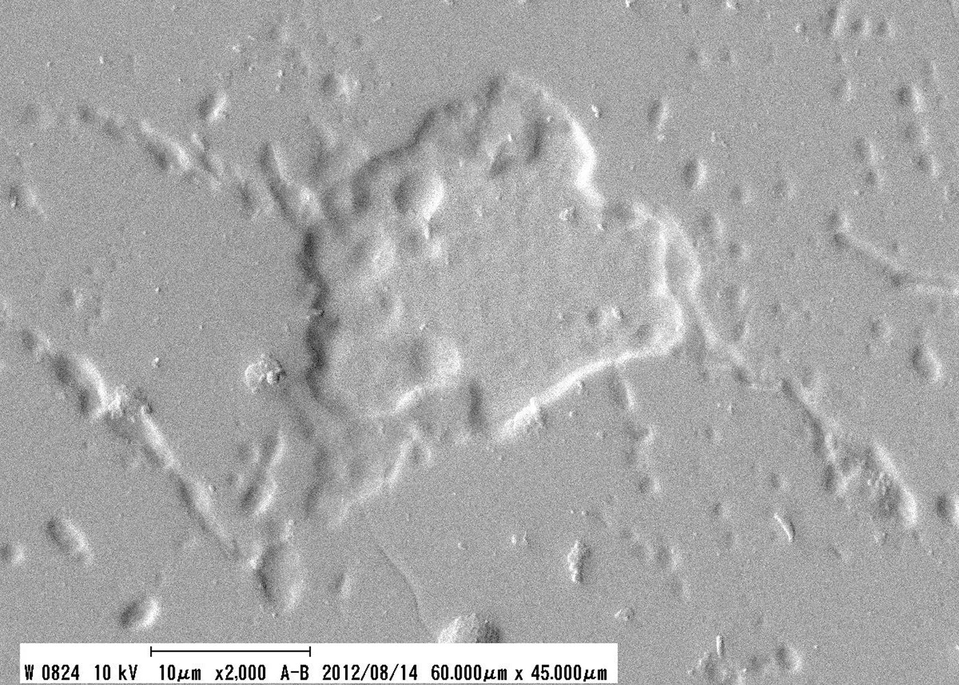
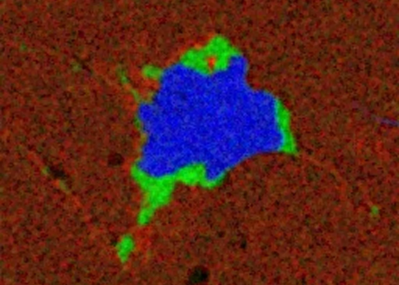
Red: Ca, Blue: S, Green: Al
Example of Expansion Panel + Joystick Installation
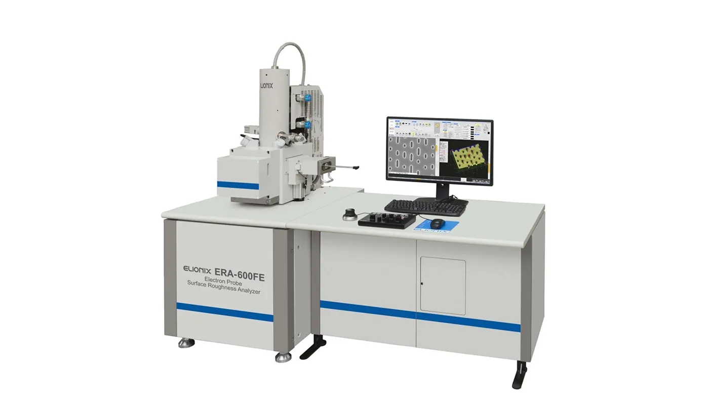
Example of EDS + EBSD Option Installation
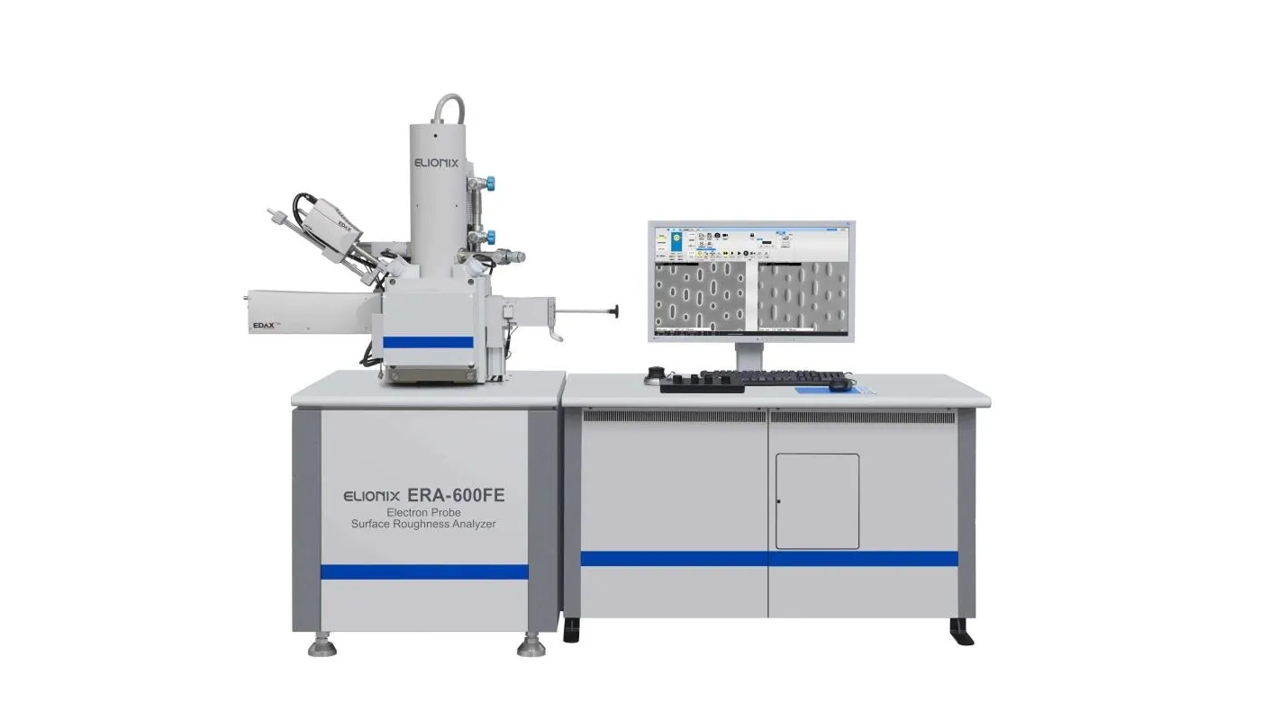
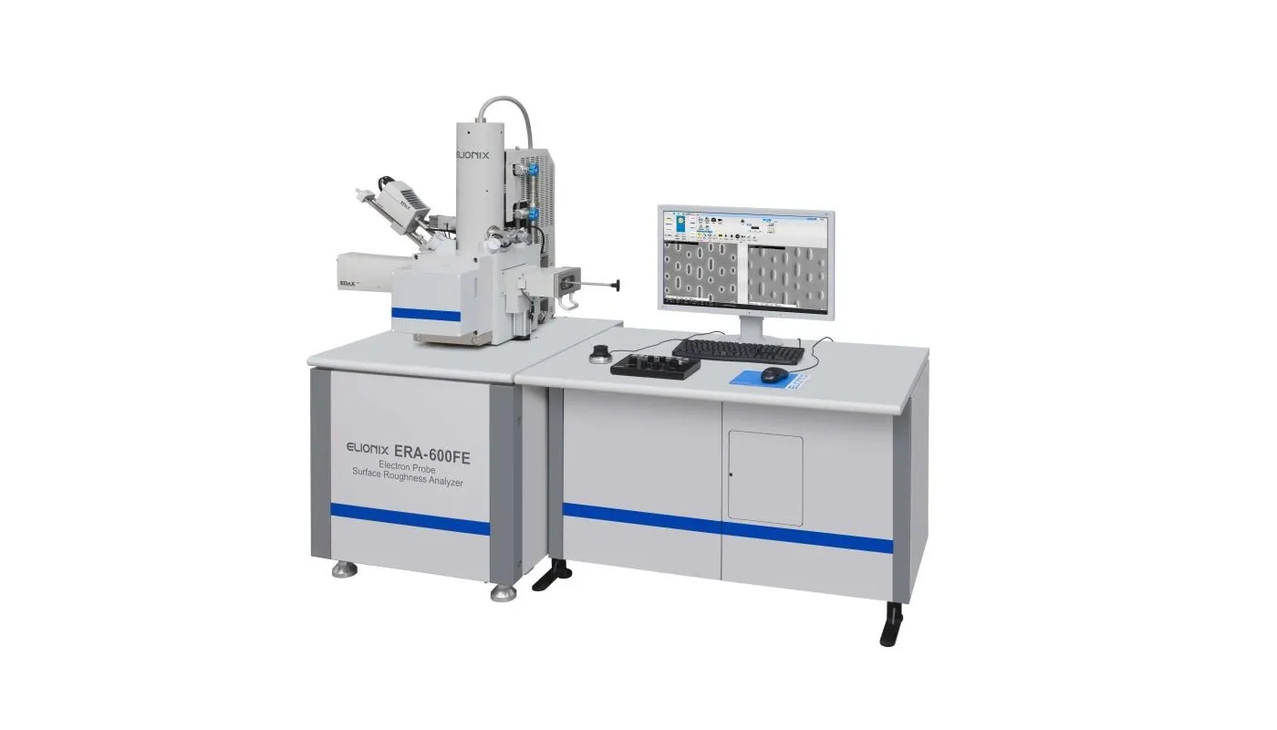
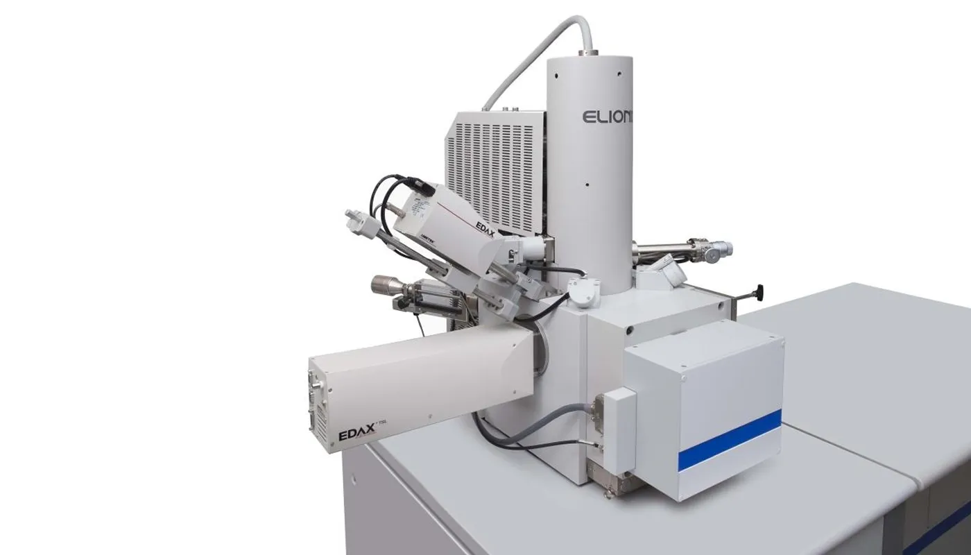
Options
ENT-ERA (SEM, EBSD)
Coordinate-Sharing Holder System
Both are instruments used for observing, analyzing, and evaluating the physical properties of specimen surfaces; by integrating them, you can obtain analytical results that significantly expand the capabilities of each individual instrument.
- This eliminates the need to remove and re-mount samples, enabling smooth sample transfer between instruments.
- Stage coordinate conversion between ERA Series, ENT-NEXUS, and ENT Series instruments is straightforward, allowing for the selection of the exact same field of view and measurement location in a matter of seconds.
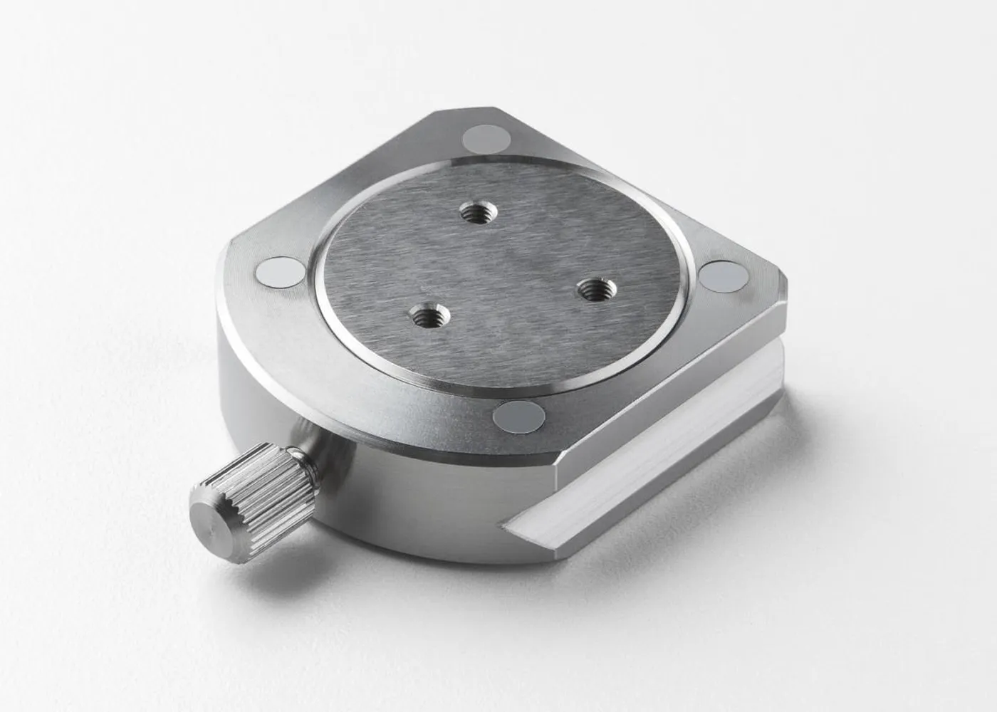
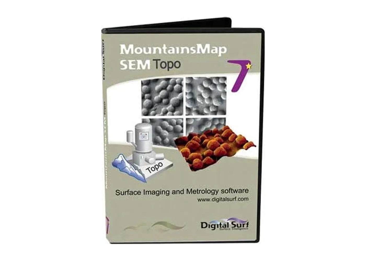
Digital Surf's MountainsMap®
Topo Software
Key Specifications
| ERA-600FE | ERA-600 | |
|---|---|---|
| Electron Gun | ZrO/W thermoelectric emission type | Precision tungsten |
| Accelerating Voltage | 0.3~30 kV | 0.3~35 kV |
| Lateral Resolution | 1.2 nm:30 kV 5 nm:1 kV |
3.5 nm:35 kV |
| Magnification | x20~600,000 Magnification | x10~400,000 Magnification |
| Sample Dimensions | Φ150 ×t15 mm(max. diameter) Φ50 ×t30 mm(max. thickness) |
|
| Sample Drive | 5-axis motor drive (X, Y, Z, R, T) | |
| Secondary Electron Detectors | 4 (a,b,c,d) | |
| Height Resolution | 1 nm | |
| Measurement Direction | X-direction, Y-direction | |
| Number of Measurement Data Points | 3D measurement data: max. 17.28 million points; 1-line measurement data: max. 32,767 points | |
| Surface Characteristics Parameter Calculation |
Bird's-eye view, contour lines, number of peaks, area fraction, grain size, surface area JIS Standard Parameters (Compliant with JIS B 0601-2001) |
|
| Options | Control panel, joystick, BSE detector, EDS, WDS, EBSD, preliminary evacuation chamber (4-inch type), navigation camera, various sample holders, Digital Surf Mountainsmap software— |
|
Download Related Materials
- Examples of Cutting Edge Evaluation of Precision Cutting Tools Using the ERA-600 and ERA-600FE
- Surface Topography Evaluation Technology Using Electron Beams Source: *Surface Technology* Vol. 57, No. 8, pp. 18–22
- Principles of the ERA-8900 and 8900FE